

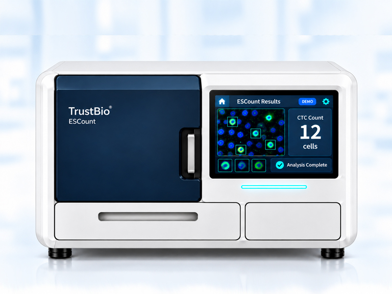
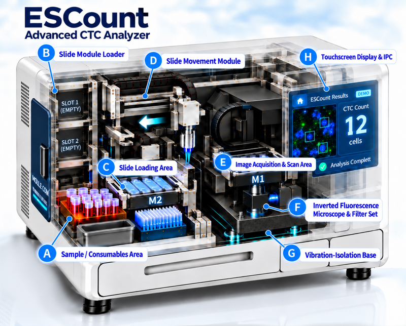
ESCount™ Product Introduction
Breaking Through the Limitations of Conventional Detection to Precisely Capture Every Critical Cell
CTC Circulating Tumor Cell Analyzer Powered by the ESCount™ Patented BioChip
By integrating proprietary antibody-independent fluorescent staining technology with AI-powered automated image analysis, ESCount™ provides a high-recovery, high-integrity liquid biopsy solution for cancer research, early cancer risk screening, post-treatment recurrence monitoring, and personalized therapy evaluation.
What Are Circulating Tumor Cells (CTCs)?
Circulating Tumor Cells (CTCs) are cancer cells that detach from primary or metastatic tumors and enter the peripheral bloodstream.
During cancer progression, CTCs serve as the "seeds" of metastasis. Even when tumors are extremely small and remain undetectable by conventional imaging techniques such as CT or MRI, CTCs may already be present in circulation. By capturing and analyzing these intact viable tumor cells, clinicians and researchers can gain real-time, dynamic insights into tumor biology and disease progression.
Why Perform CTC Testing? Who Can Benefit?
Traditional tissue biopsy is invasive, unsuitable for frequent sampling, and often limited by tumor heterogeneity. Conventional blood-based tumor markers, such as CEA and CA19-9, may also suffer from limited sensitivity and specificity.
CTC testing requires only a routine blood draw and helps fill a critical gap in precision oncology by providing accessible, minimally invasive, and clinically actionable information.
Why Choose CTC Testing?
Ultra-Early Cancer Risk Detection
Detect trace amounts of circulating cancer cells before tumors become visible through conventional imaging, even when lesions are as small as 1-2 mm.
Real-Time Disease Monitoring
The non-invasive blood-draw approach supports frequent follow-up testing and provides dynamic insights into a patient's current disease status.
Overcoming Tumor Heterogeneity
Blood-borne CTCs can represent cellular information from both primary and metastatic lesions, providing a more comprehensive tumor profile than single-site tissue biopsies.
Comprehensive Cellular Information
Unlike ctDNA assays that analyze fragmented tumor DNA, CTC analysis preserves intact cellular morphology and biological activity, enabling downstream molecular studies and drug sensitivity testing.
Target Users and Application Scenarios
High-Risk Individuals and Health-Conscious Populations
Early Cancer Risk Screening
Ideal for individuals with a family history of cancer, long-term exposure to carcinogenic risk factors such as smoking, environmental pollutants, or chronic inflammation, as well as those seeking advanced preventive health examinations.
Cancer Patients
Personalized Treatment Assessment
Regular monitoring of CTC counts before and during treatment enables rapid evaluation of chemotherapy, targeted therapy, and immunotherapy effectiveness, supporting timely optimization of treatment strategies.
Postoperative and Recovery Patients
Prognosis and Recurrence Monitoring
Assess minimal residual disease (MRD) following surgery and identify recurrence or metastasis risks before clinical symptoms or imaging evidence emerge.
Oncology and Translational Researchers
Research Applications
A powerful research tool for investigating cancer metastasis, mechanisms of drug resistance, and novel therapeutic development.
Core Technology: Patented ESCount™ BioChip
Most existing CTC detection technologies face two major limitations.
Immunomagnetic enrichment methods rely on antibody-based capture and may fail to detect highly metastatic cancer cells that have undergone epithelial-mesenchymal transition (EMT) and lost EpCAM surface expression.
Physical filtration platforms based on microfluidic channels or membrane filters depend on cell size, morphology, and deformability for separation. These approaches may miss smaller CTCs while fluid shear stress can damage fragile tumor cells during processing.
The patented ESCount™ BioChip overcomes these challenges through an innovative capture approach.
Antibody-Free Enrichment
Eliminates dependence on a single surface marker such as EpCAM, enabling comprehensive capture of heterogeneous CTC populations, including epithelial, mesenchymal (EMT), and hybrid phenotypes.
Size- and Deformability-Independent Capture
Unlike conventional microfluidic filtration systems, ESCount™ does not rely on cell size, shape, or deformability. By eliminating mechanical compression and fluid shear stress, the platform preserves the structural integrity of fragile CTCs.
Proprietary CTC-Specific Fluorescent Dye Cocktail
A unique staining formulation targeting cancer-specific metabolic activity and biomarkers enables precise discrimination between viable CTCs and normal blood cells, maximizing both recovery rate and detection accuracy.
AI-Powered Imaging and Analysis System
Combining high-resolution optical imaging with deep-learning algorithms, the system automatically identifies, localizes, and enumerates specifically stained CTCs while minimizing subjective interpretation and significantly improving throughput and reproducibility.
Four Key Advantages of ESCount™
Minimal Blood Volume Requirement
Requires only 2.5 mL of whole blood, significantly reducing the burden of blood collection, particularly for elderly and physically frail individuals.
High Recovery Rate and Exceptional Cell Integrity
By eliminating antibody-capture bias and microfluidic-induced cellular damage, ESCount™ more accurately reflects the true quantity and morphology of CTCs present in circulation.
Safe and Non-Invasive
Only a routine venous blood draw is required. No radiation exposure, surgical procedures, or recovery time are involved, enabling convenient longitudinal monitoring.
Standardized and Efficient Workflow
The combination of proprietary fluorescent staining and AI-assisted image analysis delivers objective, quantitative, and highly reproducible results through a standardized testing process.
ESCount™ Standard Workflow
From sample collection to report generation, ESCount™ offers a streamlined workflow designed for seamless integration into both clinical and laboratory environments.
Step 1. Sample Collection
Collect 2.5 mL of peripheral whole blood into a dedicated collection tube.
Step 2. Sample Preparation and BioChip Loading
Following gentle sample processing, the cell suspension is loaded onto the patented ESCount™ BioChip. The workflow avoids high-pressure microfluidic compression, ensuring minimal cellular damage.
Step 3. CTC-Specific Fluorescent Staining
The proprietary CTC-Specific Dye Cocktail selectively labels target CTCs while minimizing interference from leukocytes and other background cells.
Step 4. AI Imaging and Analysis
The BioChip is inserted into the CTC Circulating Tumor Cell Analyzer for automated multi-channel high-resolution fluorescence scanning. AI algorithms then identify cellular characteristics and accurately enumerate captured CTCs.
Step 5. Report Generation
A structured analytical report is automatically generated, including high-resolution CTC images and quantitative results to support clinical decision-making and scientific research.
